

Digital X-Rays & iTero Scanning in St. John's
Most people assume a dental visit is the same everywhere: a quick look in the mouth, some poking around with a metal instrument, and maybe a blurry film that gets clipped to a lightbox. That picture is outdated. If you are searching for a digital X-rays dentist in St. John's, it is worth understanding what modern diagnostic imaging actually does differently, because the gap between old-technology dentistry and current practice is wider than most patients realize. At Stavanger Dental, diagnostic appointments begin with two tools that together produce a far more complete picture of oral health than film-based methods ever could: digital radiography and iTero intraoral scanning.
Table of Contents
Quick Takeaways
Key Insight
Explanation
Significantly lower radiation exposure
Digital X-rays use considerably less radiation than traditional film, with studies showing reductions of 50 to 80 percent compared to conventional intraoral film methods.
Instant image availability
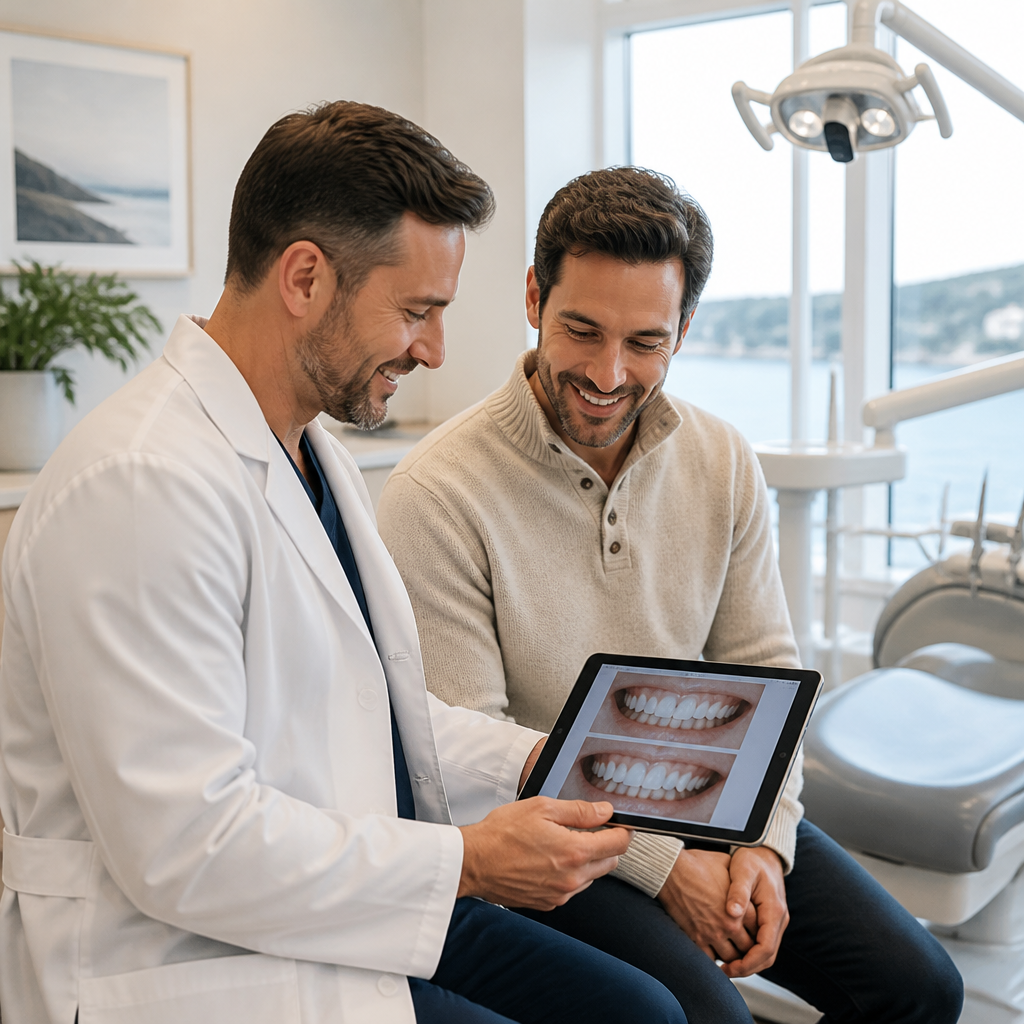
Digital radiographs appear on a chairside monitor within seconds, allowing the dentist and patient to review findings together during the same appointment.
iTero scanning replaces physical impressions
A precise 3D digital scan of the teeth and gums replaces the older tray-and-putty impression method, which many patients find uncomfortable.
Higher accuracy for restorations and orthodontics
iTero scanners are recognized by independent studies for their accuracy in restorative cases, reducing the need for remake impressions and improving fit for crowns, veneers, and aligners.
Early detection of hidden problems
Digital X-rays reveal early-stage cavities between teeth, bone loss, cysts, and issues below the gumline that a visual exam alone cannot identify.
Patient-friendly communication
Seeing a high-resolution image on a monitor makes it easier for patients to understand a diagnosis and make informed decisions about treatment.
Both tools are used at Stavanger Dental
Stavanger Dental uses both digital X-rays and iTero digital scanning as part of every comprehensive dental examination in St. John's.
What Are Digital X-Rays and How Do They Work?
Digital radiography replaces the old silver-halide film with an electronic sensor that captures X-ray data and sends it directly to a computer. The image appears on a monitor within seconds. There is no darkroom, no chemical processing, and no waiting. The dentist can zoom in, adjust contrast, and annotate findings in real time while the patient is still in the chair.
The radiation dose reduction is a genuine clinical advantage. Research published in dental radiology literature documents that digital imaging requires 50 to 80 percent less radiation exposure than conventional intraoral film-based radiography. For families bringing children in for routine checkups, that reduction matters.
Image quality also improves in ways that directly affect diagnosis. Higher sensor sensitivity means the dentist captures more detail with less exposure. Early interproximal cavities (the kind that form between teeth where a visual exam cannot reach) show up earlier. Bone density changes around tooth roots, signs of infection, and impacted teeth that are not yet symptomatic are all visible at a stage when treatment is simpler and less expensive.
Pro tip: Ask your dental team to walk you through what they are seeing on the monitor during your X-ray review. A dentist who takes five minutes to explain an image is giving you information that genuinely changes how you think about your own oral health.


Digital records are also stored electronically, which means they can be compared over time with precision. A change in bone level between this year's X-ray and last year's is measurable in a way that eyeballing two film sheets never was. That longitudinal comparison is how early-stage gum disease and slow-developing cysts get caught before they become surgical problems.
The iTero Scanner Explained
The iTero intraoral scanner is a handheld wand that captures tens of thousands of image frames per second and assembles them into a detailed 3D digital model of the teeth, gums, and bite. The process takes a few minutes and requires no trays, no impression material, and no gagging. The finished model appears on screen in real time as the dentist moves the wand around the mouth.
Why Digital Impressions Beat Traditional Putty
Traditional impressions involve filling a tray with a viscous material, pressing it against the teeth, holding it in place for several minutes, and then waiting for the material to set. Patients with a strong gag reflex, anxiety, or a small mouth often find this process difficult. Beyond comfort, the physical impression can distort slightly as it is removed from the mouth or during transport to the lab, which is why remakes happen.
Surveys of practitioners using iTero scanners have found an estimated 36 percent reduction in impression remakes compared to traditional methods. Fewer remakes mean fewer follow-up appointments and better-fitting final restorations. For a crown or a set of Invisalign aligners, that accuracy directly affects how well the appliance fits from the first delivery.
The Outcome Simulator
One feature that patients consistently find useful is the iTero Outcome Simulator. Using the scan data already captured, the software can generate a realistic projection of how teeth could look after orthodontic treatment or other cosmetic procedures. This is not a stylized rendering. It is based on actual scan geometry, so the projected result reflects realistic tooth movement and proportions. Patients considering Invisalign at Stavanger Dental can see a simulation during the same appointment as the scan.
A dental diagnosis is only as good as the information it is based on. When the information is incomplete, so is the treatment plan. Digital imaging closes that gap between what is visible and what is actually happening in the mouth.
Real Diagnostic Benefits for Patients
The most important thing digital imaging changes is not speed or convenience. It is what gets found and what gets missed. Traditional film has real limitations in resolution, storage, and the ability to enhance images after the fact. A borderline finding on film often remains a "watch and see" note in the chart. The same finding on a digital X-ray with contrast adjustment becomes something definitive enough to act on.
Finding Problems Earlier Saves Money and Treatment Time
A cavity caught at the enamel stage requires a small composite filling. The same cavity caught six months later, after it has progressed into dentin, typically requires a larger restoration. Caught a year after that, it may need a root canal and a crown. The cost and complexity multiplies at each stage. Digital X-rays make earlier detection routine, not exceptional.
The same logic applies to periodontal disease. Early bone loss is quantifiable on a digital radiograph. A dentist who can show a patient a measurable change on screen, compared to a previous image, has a far more compelling conversation about the need for more frequent cleanings or a referral to the periodontist than one relying on probing depths alone.
What Digital Imaging Finds That a Visual Exam Cannot
A clinical exam, no matter how thorough, is limited to what is visible above the gumline and between teeth from a direct line of sight. Digital X-rays reveal issues in several areas that a visual exam simply cannot reach:
Cavities forming between teeth before they break through to the surface
Bone density changes indicating early or moderate periodontal disease
Abscesses and cysts forming at the roots of teeth
Impacted wisdom teeth and their proximity to adjacent roots or the nerve
Root fractures and resorption
Changes in the jawbone over time
Pro tip: If you have not had a full set of dental X-rays in more than two years and you have changed dental providers, ask for a complete radiographic survey at your first appointment. Continuity of imaging records is one of the most overlooked aspects of preventive dental care.

Comparing Dental Imaging Approaches
Not all dental imaging is the same. The table below compares the three main approaches relevant to a general dental practice like Stavanger Dental.
Feature
Traditional Film X-Rays
Digital X-Rays
iTero 3D Intraoral Scan
Radiation exposure
Higher (baseline reference)
50 to 80% less than film
None (optical, not radiographic)
Image availability
Minutes to develop
Seconds, on-screen immediately
Real-time 3D model during scan
What it shows
Internal tooth and bone structures
Internal tooth and bone structures with enhanced detail
External tooth surfaces, gum contours, bite relationship
Patient comfort
Small sensor/film can be uncomfortable
Small sensor, similar comfort to film
No trays, no putty, minimal discomfort
Primary use
Cavity detection, bone assessment
Cavity detection, bone assessment, early disease detection
Orthodontic planning, restorations, treatment simulation
Records storage
Physical film, prone to damage
Digital, easily stored and compared over time
Digital, integrates with lab and aligner systems
Remake/error rate
Not applicable
Not applicable
Estimated 36% fewer remakes vs. traditional impressions
It is worth noting that digital X-rays and iTero scanning are complementary, not interchangeable. They answer different diagnostic questions. A digital X-ray shows what is happening inside and beneath the tooth. An iTero scan maps the surfaces and relationships between teeth. A complete diagnostic picture requires both.
How Digital Scanning Supports Invisalign and Restorations
Stavanger Dental offers Invisalign as part of its orthodontic services, and the iTero scanner is central to how that process works. When a patient is assessed for Invisalign, the scan data is submitted directly to Align Technology, which uses it to fabricate the custom aligner series. The 3D scan is more accurate than a physical impression, which means the aligners are designed from a more precise model of the patient's actual dentition.
Crowns, Veneers, and Bridges
For restorative work, the accuracy of the impression directly determines how well a crown or veneer fits at delivery. Research in restorative dentistry has shown that digital workflows produce smaller gaps and require less adjustment before cementation compared to conventional impression techniques. For patients getting a crown or bridge at Stavanger Dental, that means fewer appointments for adjustments and a better long-term fit.
The scan also integrates directly with dental laboratories. Instead of a physical impression being shipped in a box, the digital file is transmitted electronically. This reduces turnaround time and eliminates the risk of impression distortion during shipping. For patients who want restorations completed as quickly as possible, that matters in practice.
Dental Implant Planning
When dental implants are being planned, the iTero scan works in combination with the digital X-ray and, where required, cone beam CT imaging, to give the treatment team a complete map of the surgical site. Bone volume, adjacent tooth roots, and soft tissue contour all factor into implant placement decisions. Having all three data sources in digital format makes that planning more precise and communicable to any specialist involved in the case.
What to Expect at Your Appointment
At Stavanger Dental in St. John's, a comprehensive dental examination starts with digital X-rays and an iTero scan before the clinical exam begins. This sequence matters. The dentist reviews the images first, which means the hands-on examination is guided by what the diagnostics have already revealed, rather than the other way around. Findings from imaging direct where to probe, what to measure, and what questions to ask about symptoms.
The entire imaging process, X-rays and iTero scan combined, typically adds only a few minutes to a new patient appointment. The benefits of that small time investment are substantial: a baseline record that can be compared at every future visit, a plan that accounts for things a visual exam alone would miss, and a set of images you can actually see and understand on the same monitor the dentist is using.
For patients covered under the Canadian Dental Care Plan, these diagnostic services are part of the comprehensive care Stavanger Dental provides. If you are a new patient, the practice is currently welcoming families and individuals of all ages at its St. John's location.
Frequently Asked Questions
Are digital X-rays safe for children and pregnant patients?
Digital X-rays use significantly less radiation than traditional film, making them appropriate for routine use in children. For pregnant patients, dentists typically follow a conservative approach and take X-rays only when clinically necessary, with appropriate shielding. The decision is always made on a case-by-case basis with patient safety as the priority. If you have specific concerns, discuss them with your dentist before the appointment.
How often do I actually need dental X-rays?
Frequency depends on individual risk factors. Patients with a history of frequent cavities, active gum disease, or complex restorations may need X-rays more often than those with consistently healthy checkups. For a low-risk adult with good oral health, bitewing X-rays every 12 to 24 months and a full series every few years is typical. Your dentist at Stavanger Dental will recommend an interval based on your specific clinical situation, not a one-size-fits-all schedule.
Does an iTero scan replace X-rays entirely?
No. An iTero scan and digital X-rays capture different types of information. The iTero scanner maps the external surfaces of the teeth and gums in three dimensions. Digital X-rays show internal structures: the inside of teeth, roots, surrounding bone, and anything below the gumline. A complete diagnostic picture requires both. Using one without the other leaves significant information gaps.
Is the iTero scanning process uncomfortable?
For most patients, an iTero scan is considerably more comfortable than a traditional putty impression. There is no tray being pressed against the gums and no material to hold in place while it sets. The handheld wand moves around the mouth without applying significant pressure. Patients with a strong gag reflex, in particular, tend to find the digital scan much more manageable than conventional methods.
Can I use digital scan results for Invisalign at Stavanger Dental?
Yes. Stavanger Dental uses iTero scanning as part of the Invisalign assessment and treatment process. The 3D scan is submitted directly to Align Technology to design your custom aligner series. The Outcome Simulator feature can also show you a projected treatment result during your consultation, so you have a realistic visual reference before you commit to treatment.
What if I need to transfer my dental records to another provider?
Digital records, including X-rays and iTero scan files, are stored electronically and can be transferred to another provider more easily and accurately than physical film or plaster models. When you request your records, you receive the same high-quality images the dentist used, not a photocopy of a film. This continuity of imaging data benefits your care no matter where you are treated in the future.
If you have questions about the diagnostic technology used at your next appointment, or if you want to share your own experience with digital dental imaging, we would be glad to hear from you.
We would love your feedback and any insights you would share with others. What perspective would you add?


Comments